It was 1910 and department chair Emil Holmgren was pleased: the Karolinska Instituteâs Department of Histology had inaugurated new, state-of-the-art premises, with enlarged spaces for teaching and research. The new laboratories all had sinks with running water, ventilation, and cabinets for chemicals and optical instruments, thermometers, microtomes, and other necessary equipment. New microscopes were installed for students and scientists, and separate labs set up for the teaching assistant, the specimen preparator, and the department chair. There was a library, a room for wall charts, photomicrography equipment, and a good many cabinets for microscopy specimens mounted on glass slides. The embryological teaching collection contained slide series of fish, amphibian, bird, and mammal embryos in different stages of development. Complete series also existed of human embryos from the third, fourth, and fifth weeks, as well as series from later stages of development.1 The new facilities and their equipment displayed a commitment to an empirical research tradition that had a long history at the Karolinska Institute (KI).
Embryology was a vital field of medical scientific research and teaching at the time, in Sweden as well as internationally. The field included studies of human and animal gametes, fertilization, and embryonal and fetal development (normal as well as pathological). Like other branches of biological science, it depended on developing technologies and methods of observation and circulation. This chapter will explore and discuss material and visual aspects of research on the early stages of life, covering human as well as animal embryology and fetal anatomy. It will investigate the work of a few scientists at the KI. First, prominent rector and anatomy chair Anders Retzius, who ushered in a new era in Swedish anatomical study (including comparative, microscopic, and pathological anatomy), based on empirical observation. Second, his son Gustaf Retzius, a leading figure in histology, neuroanatomy, and physical
The KI was founded in 1810, ostensibly as a school for field surgeons. But its leading men aimed at building an institution for combined medical and surgical education, founded on the principles of scientific researchâa novelty within higher education at the time.3 Throughout the nineteenth century, KI professors were striving for a position as Swedenâs leading medical school and research institute, in constant competition with the medical faculties of the old universities at Lund and Uppsala. While the medical faculty at Uppsala long adhered to an idealist tradition, KI professors promoted medicine founded on empirical science and clinical practice. KI scientists thus created a productive milieu that became a driving force in the renewal of Swedish medical training and a dynamic node in the transnational circulation of knowledge. They learned from, applied, and developed approaches from the Paris school of anatomy, with its focus on hands-on experience, pathology, and morphological comparison across species. But, beginning in the 1820s, attention shifted to the German lands, where the most interesting research in microscopic anatomy was happening.4 The Nordic networks of scientific exchange were already strong, but were further strengthened during the nineteenth century, with the start of regular conferences in 1839.5 KI scientists participated in and contributed to the transnational scientific exchange through publishing internationally (mostly in German, but also in French and English); presenting
Research on embryos and fetuses was a process of visualization and materialization: observation, analysis, and communication depended on creating objects to study by applying a multitude of techniques, instruments, and chemicals. Embryonic specimensâonce received in the anatomy museum or histology labâhad to be stabilized, cut, colored, mounted on glass slides, suitably lit, and enlarged in order to be studied and depicted. Drawings and, from around 1900, photomicrographs were integral to the research process, and producing images a primary concern for the scientists. Plaster casts, wax models, and stereoscopic photography gave stable three-dimensional form to fragile embryonic and fetal bodies. Therefore, studying visualizations is key to understanding anatomical (including embryological) knowledge production in history. Published images were not just complementary to written accounts of research findings: making images was an integral part of the research process, and the resulting visuals could be as valuable as textual descriptions and arguments (if not more). For this reason, I prefer to call them visualizations rather than âillustrations,â which would indicate that the images were secondary to the words. Visualizations were stand-ins for specimens, and were necessary to communicate research findings, facilitating circulation and remote witnessing of the museum specimens. Specimen collections, in turn, functioned as libraries of existing research at the same time as they offered material for new studies.8 A well-functioning university department needed a high-quality and up-to-date infrastructure of equipment for research and teaching. Published research articles, monographs, and textbooks included detailed discussions of methods and technologies used, and reflections on the epistemic merits of their useâwhat Jutta Schickore has called âsecond-order,â or reflexive, research concerns.9 I will analyze visual and material aspects of the research process by focusing on such reflections on methods and technologies,
1 Mammalian Eggs, Embryonal Gills, and Fetal Malformations
Embryology appeared as a focused scientific pursuit around the turn of the nineteenth century, when comparative anatomists, physiologists, and zoologists began a systematic analysis of developing embryos, reaching for an understanding of the earliest stages of life.10 Whereas nineteenth-century zoologists mainly focused on invertebrates, vertebrate embryology was studied in the departments of anatomy, histology, and physiology at medical faculties and institutes. But even though embryology became an essential part of anatomy and zoology curricula, and a central field of biological research, it did not become a unified discipline, with its own departments, chairs, and journals.11 At the KI, human embryos and fetuses were studied at the anatomy department, in comparison with other mammals. But embryos and fetuses also showed up in research and curricula in histology (microscopic anatomy), pathological anatomy, gynecology and obstetrics, physiology, and forensic medicine.12
Anders Retzius established embryonic and fetal anatomy as part of the medical studentsâ anatomy curriculum in the 1820s. During his time as professor of anatomy at the KI (from 1824 until his death in 1860), he also researched normal and pathological embryology, introduced new findings and literature in the field to the readers of the proceedings of the Swedish Society of Medicine, and prepared a multitude of fetal specimens for the KIâs anatomy museum.13 And the topic of embryology became an important part of the effort to make medicine more scientific, as did microscopic anatomy in general.
On his first trip abroad in 1828, Anders Retzius traveled to Berlin, to attend the meeting of the Society of German Natural Scientists and Physicians, convened by the Humboldt brothers at Berlin University. The European continent was opening up after the Napoleonic Wars: traveling, correspondence, and the circulation of print material became easier, which made it possible for scientists to expand their networks and communicate in new ways. In Berlin, Retzius connected with a cohort of young men who were reshaping the life sciences at
Just like Johannes Müller and other post-Romantic German colleagues, Retzius was committed to careful empirical study, and skeptical of speculation. The new achromatic microscopy lenses of the 1820s and 1830s made it possible to study tissues at greater resolutions than before, down to the cellular level. And the new embryology depended on these instruments of observation.17 The excitement about embryology went hand in hand with this development of new technologies. But the focus of study was not just human
One central goal of embryological studies at this time was to distinguish abnormal forms of development by studying structural abnormalities, stillbirths, and other unusual manifestations of generation and fetal development. Case reports of unusual births were common in journals and meetings throughout the nineteenth century, although their character changed over time. In the first half of the century, many accounts of malformed fetuses were based on the maternal impression theory, which connected malformations to events during pregnancy. As detailed studies on embryonal and fetal development accumulated over the years, the collective body of knowledge increased, and the questions regarding malformations became more precise. What, exactly, was it that went wrong in the developmental process, when a baby was born with a specific malformation? Explanations began to be sought more often in the process of fetal development than in the motherâs experiences during gestation.18
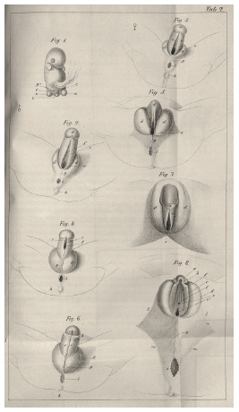
Anders Retzius took an interest in both normal and abnormal fetal development, and reported on the development of genitalia as early as 1828.19 Retzius prepared specimens of normal and abnormal embryos and fetuses for the KIâs anatomy museum, and put them on display alongside wet and dry normal and pathological specimens of adult humans, a growing series of racial crania, and a large number of animal skeletons and preparations. The KIâs professors and affiliated physicians collected fetal specimens in their practices and clinics. But specimens could also be sent to the KI by provincial physicians hoping to make a name for themselves and strengthen their position within contemporary medical networks. The museum thus became a network hub, with Anders Retzius at its center. The trade in specimens helped establish and uphold relationships and hierarchies within the community of medical doctors. Published case accounts and catalogue entries mentioned donorsâ names and gave them a recognized place in the scientific network (although the KIâs museum catalogues were lost in a fire in 1892).20 When Retzius received embryonic and fetal
Retziusâs museum-based, comparative research method was an analytical and visual way of knowing. The visualizing work started at the dissection table and went on to materialize specimens for research and display. For early nineteenth-century embryology, this method was perfect. Creating series of specimens helped scientists understand what was normal development and what was not. Comparative analysis was necessary and depended

Schematic drawings of genital development in human embryos of both sexes, from specimens in the KIâs Anatomy Museum. Lithographic prints in Anders Retzius, âHändelse af Hypospadi,â 9, Tab. 2. Hagströmer Library, KI.
2 Comparative Studies of Fetal Development: Brains and Proportions
Although Anders Retzius was a very productive researcher and author of research papers, he very rarely theorized or made statements of scientific principles. And he never published comprehensive monographs or atlases, preferring active research work to writing. In a rare exception to his usual empirical focus, however, Retzius ventured a theoretical approach to brain development in an 1844 lecture at the Royal Swedish Academy of Sciences. Referring again to the recapitulation theory, he claimed that human fetal brain development goes through the shapes of the âlower classes of animals.â This was a ânatural lawâ described by Friedrich Tiedemann, who had also shown that the brain hemispheres developed from front to back in his âclassicalâ work on the fetal development of the human brain from 1816. How to understand and locate the higher functions of the brain was, according to Retzius, âone of the greatest problems of our time.â Retzius connected the theoretical perspective to his own embryo specimens in the KI museum, and showed drawings of these to the audience. The printed lecture is instead accompanied by four diagrammatic representations of human brain development, which visualize the theory rather than actual brain specimens.24
While Anders Retzius dominated Swedish anatomy from around 1830 until 1860, his son Gustaf Retzius was the most prolific Swedish researcher in the anatomical sciences from 1876 to the mid-1910s. Continuing in his fatherâs footsteps, Gustaf studied a wide range of subjects in comparative anatomy,
Diagrammatic representation of fetal brains. Lithographic prints in Anders Retzius, âOm bildningen,â Tab. B. Hagströmer Library, KI.
One of Gustaf Retziusâs main research interests was comparative brain anatomy: morphological and macroscopic, as well as microscopic. Methods of preparation were crucial to this study, and Retziusâs publications always reflect on methodology and âsecond-orderâ research questions in great detail. One lecture, presented to the Swedish Society of Biology in 1889, is particularly noteworthy in this regard. It is entirely devoted to brain preparation and accompanied by demonstrations of specimens and drawings. âA badly preserved brain is of little value,â Retzius asserts in the opening sentence of the printed talk. And he devotes a section to fetal specimens, comparing various techniques and fluids for fixation and preservation of this delicate organ. For
In 1896, Retzius published a book on the macroscopic anatomy of the human brain, Das Menschenhirn, which contained case studies of adult and fetal brains in two oversize tomes. The text in the first volume is mostly descriptive, containing few and modest conclusions: womenâs brains did not differ from menâs brains in any significant way, even if they were generally smaller, less complex, and displayed a tendency to be lighter, which could possibly influence creativity and intellectual processing. Retzius came to a similar conclusion regarding fetal brain developmentâhe had not been able to see any differences between male and female brain development. And in a typically cautious manner, he deferred to the need for more comparative studies, which should also take into account possible racial differences.28 Gustaf Retzius seemed to believe that future studies would, or could, reveal such differences in the structure of brains, but, on the basis of his own meticulously empirical research, he could not agree with those scientists who had made strong claims that placed white men in an exceptional category of neurological refinement.29
Many of the plates in Das Menschenhirn are full-size photographs of adult brains, hand printed in collotype on expensive paper by printer G. Tholander. Retzius only worked with greatly skilled collaborators, demanding the highest quality every step of the way: from his own preparations to the photographs by Christian Westphal and the drawings by medical artists Sigrid Andersson-Rissler, Hilma Bundsen, Ebba Flodman, and Gustaf Wennman.30 Whereas adult brain morphology is represented in full-size photographic reproduction,
Eight years later, Retzius published a study on the proportions of human fetuses, with a particular focus on extremities. He again collaborated with artist Gustaf Wennman and photographer Christian Westphal, who this time
Fetal brains and spinal cords produced by photographer Christian Westphal, together with artists and technicians. Collotype plate in Gustaf Retzius, Das Menschenhirn, Taf. XIV. Hagströmer Library, KI.
The topic of fetal proportions needs further study, Retzius claims, and goes on to describe in detail the relative size and length of fetuses and their arms and legs, from the second to the third month of gestation. The text is complemented by tables of measurements, taken from skeletonized fetuses, wet specimens, and some fresh, unprepared specimens. When this study was published, Retzius was no longer officially affiliated with the KI, but the text makes clear that he was still very well connected with its faculty. One series of fetuses in the study consists of thirty-nine skeletons, which had been preserved in a mixture of water, glycerin, and alcohol by a preparator credited as Mr. Roth, when Retzius was chair of the anatomy department and director of the museum of anatomy, in 1888â89. Another series had whole, undissected fetuses, which were conserved in various fluids. He had received most of these fetuses after 1890, from his colleagues at the KI, including the current museum director, anatomy chair Erik Müller. Some of the specimens belonged to the Anatomy Museum, but most of them were in Retziusâs own possession.35
Gustaf Retziusâs meticulous care in procuring, dissecting, preparing, studying, and visualizing the specimens was a method in and of itself. The goal of his comparative anatomical research was often to study large series of particular
Pencil drawings of fetal hands and feet by Gustaf Wennman. Lithographic print in Gustaf Retzius, Biologische Untersuchungen, Taf. XXIV. Hagströmer Library, KI.
3 Variations on a Theme: Sperm, Ova, and Chromosomes
Around the same time as the study of fetal proportions, Retzius began collecting, visualizing, and analyzing sperm and egg cells from humans and animals. Detailed investigations of gametes were, he believed, necessary in order to understand the process of fertilization. In 1902, he published a couple of articles on the subject of sperm morphology, following up on an earlier paper from 1881. These were followed by another fifty papers on the subject in his own publication series, Biologische Untersuchungen, the last of which was published posthumously in 1921.36 All of them contained lithographic prints from his own drawings, more than three thousand figures in all. Retzius made these precise drawings at his apochromatic Zeiss microscope, observing the specimens, which were usually fixed with osmium tetroxide or Zenkerâs fixative, preferably with sunlight as his light source. As always, he carefully described the methods and materials he used. Judging from oversized wall charts preserved in the archive of the Royal Swedish Academy of Sciences, Retzius also gave lectures on the subject of sperm morphology. In a plate from 1912, human sperm cells are lined up next to sperm of other primates to facilitate visual comparison.
Gustaf Retzius managed to obtain specimens from more than four hundred different species from all over the world, covering sperm of every type of organism, from primitive marine gastropods to apes and humans. He purchased fresh or prepared specimens (testicles or whole animals) from dealers and hunters or received gifts and donations from scientists, curators, and collectors within his immense international scientific network. Some of the animals came from zoos or museums; others were killed in the wild. Some were rare, such as the Australian echidna and Japanese giant salamander. Othersâsuch as mussels, clams, sea worms, starfish, and jellyfishâRetzius fished out of the waters near
Gustaf Retziusâs own drawings of primate sperm cells. Lithographic plate in Retzius, Biologische Untersuchungen, Taf. XVI. Hagströmer Library, KI.
Retzius also devoted time to studying ova, as well as the first phases of cell division after fertilization. Volumes 16, 17, and 18 of Biologische Untersuchungen contain a number of papers on the subject, based on careful preparation, staining, and analysis. In the introduction to three papers published in 1911, Retzius writes that the investigations are the results of his efforts to find a method that would stain the chromosomes of egg cells and sperm cells in different colors, so that he would be able to study the process of development immediately following fertilization. In Echinoderms (starfish, sea urchins, and the like), he had found that a particular staining method made the chromosomes in the nucleus of sperm cells retain a green color after entering the egg cell, whereas the ovumâs chromosomes stood out in red. This method could not easily be used in mammals, however, and this paper is devoted entirely to discussing methods of fixating, staining, and studying these minuscule parts of the cell nuclei of invertebrates. Hoping to solve some of the problems of fertilization, Retzius turned to studying fertilized eggs of the nematode Ascaris megalocephala, an intestinal parasite of horses and an âold, well-known ideal object for chromosome research.â He found the quality of the images in his âesteemed colleaguesâ published research on the subject unsatisfactory and set out to produce something superior. The Ascaris was a popular research subject because it had only two chromosomes in its cells and was easy to study. It was not easy to acquire the necessary specimens, though. Retzius failed to find worms at horse slaughterhouses in Stockholm and then turned to his colleagues Theodor Boveri and Friedrich Mevesâleading researchers on the subjectâto ask for specimens, which they willingly sent. He found these unusable because of the effect of injected dyes, and so was back to square one. Finally, he received some fresh specimens from a colleague at the KI, Emil Holmström, and could start the research.38 In the foreword as well as in his autobiography, Retzius acknowledges technician Anna Edman, who masterfully âperformed the difficult and time-consuming work of embedding, microtomizing [slicing], coloring, and mounting [the ova specimens] in Canada turpentine.â39
Gustaf Retziusâ own drawings of colored specimens visualizing the first stages of cell division in fertilized ova of Ascaris megalocephala. Chromolithographic plate in Retzius, Biologische Untersuchungen, Taf. IX. Hagströmer Library, KI.
Gustaf Retzius learned, and further developed, the latest methods of fixating and coloring microscopic specimens, which he also discussed in detail in his publications. He used the best available microscopes and printing technology.43 The outcome of his studies has the character of âmuseum scienceâ: a collecting
When Erik Müller gave his inaugural lecture as chair of histology at the KI in 1896, he also dealt with chromosomes, but in the setting of a discussion of scientific studies and theories of generation, from the eighteenth century on. Müller, who had studied with Oscar Hertwig and Wilhelm His, stressed the importance of microscopic technology, and favored âthe most exact of all methods of research, [â¦] experiment.â While his arguments regarding evolutionists and epigeneticists in his own era are obscure, his opinion on the importance of chromosomes is clear: âthe most important substance in all living organisms is the so-called chromatin.â Fertilization is a process through which nuclear parts of the sperm cell are introduced into the egg cell, Müller continues, connecting the chromatin from the father and the mother. Chromatin is what makes up the chromosomes, and it carries the inherited characteristics from the parents and start the ontogenetic development of the individual. Supporting Oscar Hertwig and Wilhelm Roux, and criticizing Ernst Haeckel and his biogenetic law (âontogeny recapitulates phylogenyâ), Müller sees cellular science as the foundation of modern biology. He ends his talk by hailing recent research findings in histology and embryology, and their influence on medical practice. Although a new approach to development and heredity was underway, Müller still emphasized the foundational character of microscopic anatomy, for both the scientist and the practicing physician.46
4 Microscopic Slides and Plaster Casts: New Approaches to Embryology
Much as the conditions of science and society had changed between the 1820s of Anders Retzius and the early 1900s of Erik Müller and Emil Holmgren, one theme connects their work at the KI: the collection, preparation, study, and
Dominant as it was in embryology, microscopic study was not suitable for all research topics. The method of observation and visualization had to be adapted to the research question. In a monograph from 1896, Erik Müller studied fetal development of abdominal organs, particularly the small intestine. Müller spent the spring and summer researching this topic, first in Stockholm, and then in Leipzig, at Wilhelm Hisâs anatomical department. He acknowledges His in the introduction, along with the sculptor Franz Josef Steger, who had taught him to make plaster casts. Steger and His had collaborated to make durable plaster models for anatomical instruction, and plaster was used in many scientific and cultural settings at the time, including anatomy: to produce molds for wax moulages and models, and to make death and life masks, anthropological casts, and copies of sculptures, portrait busts, architectural details, and archaeological objects.49 The method Müller used to study the fetuses was unusual, however. After injecting fetuses with a formalin solution through the carotid artery, and then hardening them suspended in formalin, Müller dissected the fetuses using a method developed by His, removing tissues and organs layer by
The ten image plates of the article show a combination of lithographed drawings and photographs of the plaster casts, thus visualizing a series of progressively larger fetal specimens, from five centimeters long up to fifty-five centimeters. Plate IV (figure 4.7) also shows the gradual dissection of two fetuses, by laying out a series of plaster casts next to each other. By applying this method, Müller claims to have been able to better examine and visualize the topography of abdominal organs of the fetus âin situ,â compared to studies that, for example, used sections made from frozen specimens. Müllerâs conclusion ends up comparing his own results with publications of his peers, and the methodological discussion is as crucial here as it ever was for Anders and Gustaf Retzius.51
Photograph of a series of plaster casts displaying progressive dissections of the intestines of two fetuses, seventeen and twenty centimeters long. Photo-lithographic plate in Müller, âBeiträge zur Anatomie,â Taf. IV. Hagströmer Library, KI.
Photomicrographs of the first stages of cell division in a fertilized ovum of Ascaris megalocephala, produced at the Department of Histology at the KI. Halftone print in Holmgren, Lärobok i histologi, 91, fig. 34. Hagströmer Library, KI.
Emil Holmgren, who proudly described the modern equipment of the histology department of the KI in 1910, made use of the laboratory equipment for teaching and research, but also used its photographic studio for publishing a textbook in histology in 1920.53 This volume contained 742 reproduced photographs of microscopic specimens in the departmentâs collections. Photographic micrography (not to be confused with micro-photography; i.e., minute photographs), was a technique to take images of specimens through the lens of a microscope. It had by this time become the preferred, and most modern, form for visualizing, reproducing, and circulating histological knowledge.
5 Conclusion: the Embryo as Model of Development
Between Anders Retziusâs first dissections of sheep embryos to study their gill slits and Emil Holmgrenâs handbook in histology lies a dramatic century of societal transformation. Sweden went from a poor and war-ridden country to a modern industrial state. Science and technology were at the heart of this nation-building, modernizing effort. The KI established itself as a modern research institution and medical school with high standards, dedicated to the empirical study of medicine and life science. During the nineteenth century, research in anatomyâmedicineâs foundational scienceâbecame increasingly based on comparative methods and microscopic investigations. Embryology as a subject for research and teaching was at the center of this development.55 The KIâs professors of anatomy and histology were enthusiastically teaching the latest in embryology, as well as pursuing research and participating in international networks of scientific exchange.
Scientific knowledge about generation and embryonal development increased tremendously during the nineteenth century, following von Baerâs description of the mammalian ova, Rathkeâs demonstration of fetal gill slits, and the establishment of the cell theory in the 1830s. Life scientists found the embryological field of inquiry central to their pursuit of knowledge. This had deeper connotations than mere delight in novel research findings or individual contributions that expanded the general body of knowledge. Embryonal development had taken on a symbolic meaning. Whether or not an individual scientist supported the recapitulation theoryâas formulated by Ernst Haeckel or earlier by Johann Friedrich Meckel and his contemporariesâembryology described a development from the simple to the complex, from the general
What I have focused on here, however, is not the grand theories of life and evolution, but practices of embryonic and fetal knowledge production at the KI, the work that scientists performed in their laboratories and transferred to the pages of their publications. This work was inherently visual and material. Studying, analyzing, and comparing gametes, fertilization, or embryonic and fetal development in different species required a chain of practices of collection and preparation. They involved technologies of staining, cutting, slicing, and magnifying. Artists, hunters, doctors, janitors, photographers, instrument makers, chemists, technicians, and printers were necessary collaborators in this pursuit. Specimens, which grounded the scientific value of the study, remained in the collections of the anatomy and histology departments. As I have shown, the end results were not just scientific texts, but, more importantly, visualizations based on observations of specimens, thus reflecting the process of knowledge productionâa visual and material way of knowing.
Acknowledgements
I wish to thank Michael Sappol, Carrie Greenwood, and Anna Lantz for their expertise and support.
Bibliography
Afzelius, Björn A. âGustaf Retzius and Spermatology.â International Journal of Developmental Biology 39, no. 5 (1995): 675â85.
à hrén, Eva. âFiguring Things Out: Visualizations in the Work of Anders and Gustaf Retzius, 1829â1921.â Nuncius: Journal of the Material and Visual History of Science 32, no. 1 (2017): 166â211.
à hrén, Eva. âMaking Space for Specimens: The Museums of the Karolinska Institute, Stockholm.â In Medical Museums: Past, Present, Future, edited by Samuel J. M. M. Alberti and Elizabeth Hallam, 102â15. London: Royal College of Surgeons of England, 2013.
à hrén, Eva. âMuseerna: Vetenskap i tre dimensioner.â In Johannisson, Nilsson, and Qvarsell, Medicinen blir till vetenskap, 126â69.
Alberti, Samuel J. M. M. Morbid Curiosities: Medical Museums in Nineteenth-Century Britain. Oxford: Oxford University Press, 2011.
Al-Gailani, Salim. âThe âIce Ageâ in Anatomy and Obstetrics: Hand and Eye in the Promotion of Frozen Sections around 1900.â Bulletin of the History of Medicine 90 (2016): 611â42.
Anders Retzius bref till A. H. Florman: Ett bidrag till medicinens historia i Sverige. Edited by Carl M. Fürst. Lund: Gleerups, 1896.
Baer, Karl Ernst von. Ãber Entwicklungsgeschichte der Thiere: Beobachtung und Reflexion. Vol. 1. Königsberg: Bornträger, 1828.
Bondestam, Maja. Tvåkönad: Studier i den svenska hermafroditens historia. Nora: Nya Doxa, 2010.
Bondestam, Maja. See also Larsson, Maja.
Buklijas, Tatjana, and Nick Hopwood. Making Visible Embryos (online exhibition). 2008â10. Accessed December 4, 2020. http://www.hps.cam.ac.uk/visibleembryos/.
Chorell, Torbjörn Gustafsson. âNeurovetenskap: Hjärna och psyke.â In Johannisson, Nilsson, and Qvarsell, Medicinen blir till vetenskap, 238â26.
Churchill, Frederick B. August Weismann: Development, Heredity, and Evolution. Cambridge, MA: Harvard University Press, 2015.
Churchill, Frederick B. âFrom Heredity Theory to Vererbung: The Transmission Problem, 1850â1915.â Isis 78 (1987): 337â64.
Cornwall, Jon, and Chris Smith. âAnatomical Models by F. J. Steger (1845â1938): The University of Otago Collection.â European Journal of Anatomy 18, no. 3 (2014): 209â11.
Daston, Lorraine, and Peter Galison. âThe Image of Objectivity.â Representations 40 (Fall 1992): 81â128.
Daston, Lorraine, and Peter Galison. Objectivity. New York: Zone Books, 2007.
Eriksson, Nils. âI andans kraft, pÃ¥ sannings strÃ¥tâ¦â: De skandinaviska naturforskarmötena 1839â1936. Göteborg: Göteborgs universitet, 1991.
Fiorentini, Erna. âInduction of Visibility: Reflections on Histological Slides, Drawing Visual Hypotheses and Aesthetic-Epistemic Actions.â History and Philosophy of the Life Sciences 35 (2013): 379â94.
Fleck, Ludwik. Genesis and Development of a Scientific Fact. Translated by F. Bradley and T. J. Trenn. Chicago: University of Chicago Press, 1979.
Franzén, Helena. âAtt lära sig se embryologiskt: Samlingsobjekt i forskning och pedagogiska kontexter 1870â1920.â Scandia 89, no. 2 (2023): 201â229.
Gascoigne, Bamber. How to Identify Prints: A Complete Guide to Manual and Mechanical Processes from Woodcut to Inkjet, 2nd ed. London: Thames & Hudson, 2004.
Gustafson, Tony. âAnatomi: Praktik och pedagogik.â In Johannisson, Nilsson, and Qvarsell, Medicinen blir till vetenskap, 170â99.
Gustafsson, Torbjörn. âEn harmonisk och hierarkisk ordning: Synen pÃ¥ utveckling i 1800-talets svenska biologi.â Lychnos (1994): 87â113.
Hallam, Elizabeth. Anatomy Museum: Death and the Body Displayed. London: Reaktion Books, 2016.
Holmgren, Emil. âHistologiska institutionen.â In Karolinska mediko-kirurgiska institutets historia, 3:462â82. Stockholm: Karolinska mediko-kirurgiska institutet, 1910.
Holmgren, Emil. Lärobok i histologi. Stockholm: Norstedts, 1920.
Hopwood, Nick. âEmbryology.â In The Cambridge History of Science, vol. 2, edited by Peter J. Bowler and John V. Pickstone, 285â315. Cambridge: Cambridge University Press, 2009.
Hopwood, Nick. Embryos in Wax: Models from the Ziegler Studio. Cambridge: Whipple Museum of the History of Science, 2002.
Hopwood, Nick. Haeckelâs Embryos: Images, Evolution, and Fraud. Chicago: University of Chicago Press, 2015.
Hopwood, Nick. âPlastic Publishing in Embryology.â In Models: The Third Dimension of Science, edited by Soraya de Chadarevian and Nick Hopwood, 170â206. Stanford, CA: Stanford University Press, 2004.
Hopwood, Nick. âProducing Development: The Anatomy of Human Embryos and the Norms of Wilhelm His.â Bulletin of the History of Medicine 74, no. 1 (2000): 29â79.
Hopwood, Nick, Rebecca Flemming, and Lauren Kassel, eds. Reproduction: Antiquity to the Present Day. Cambridge: Cambridge University Press, 2018.
Hopwood, Nick, Peter Murray Jones, Lauren Kassel, and James Secord. âIntroduction: Communicating Reproduction.â Bulletin of the History of Medicine 89, no. 3 (Fall 2015): 379â404.
Hopwood, Nick, Simon Schaffer, and Jim Secord. âSeriality and Scientific Objects in the Nineteenth Century.â History of Science 48, no. 3â4 (2010): 251â85.
Johannisson, Karin, Ingemar Nilsson, and Roger Qvarsell, eds. Medicinen blir till vetenskap: Karolinska Institutet under två århundraden. Solna: Karolinska University Press, 2010.
Karolinska mediko-kirurgiska institutets historia. Vols. 1â3. Stockholm: Karolinska mediko-kirurgiska institutet, 1910.
Kungl. Svenska vetenskapsakademiens handlingar 49 (1912).
Larsson, Maja. âFrom Frightening Beast to Primitive Stage: On the Normalization of the Monstrous Body in Swedish Medicine.â In Body Claims, edited by Janne Bromseth, Lisa Folkmarson Käll, and Katarina Mattsson, 26â57. Uppsala: Uppsala University, 2009.
Larsson, Maja. See also Bondestam, Maja.
Latour, Bruno. âDrawing Things Together.â In Representation in Scientific Practice, edited by Michael Lynch and Steve Woolgar, 19â68. Cambridge, MA: MIT Press, 1990.
Lenoir, Timothy. The Strategy of Life: Teleology and Mechanics in Nineteenth-Century German Biology. Dordrecht: Reidel, 1982.
Lindblad, Tomas. Gustaf Retzius: A Biography. Stockholm: Hagströmerbiblioteket, 2007.
Ljungström, Olof. Oscariansk antropologi: Etnografi, förhistoria och rasforskning under sent 1800-tal. Hedemora: Gidlunds, 2004.
Lucae, Johann Christian Gustav. Zur organischen Formenlehre. Frankfurt am Main: Varrentrapp, 1844.
McLeary, Erin Hunter. âScience in a Bottle: The Medical Museum in North America, 1860â1940.â PhD diss., University of Pennsylvania, 2001.
Morgan, Lynn M. Icons of Life: A Cultural History of Human Embryos. Berkeley: University of California Press, 2009.
Müller, Erik. âAnatomiska institutionen.â In Karolinska mediko-kirurgiska institutets historia, 3:69â138. Stockholm: Karolinska mediko-kirurgiska institutet, 1910.
Müller, Erik âBeiträge zur Anatomie des menschlichen Foetus.â Kongl. Svenska Vetenskaps-akademiens handlingar 29, no. 2 (1897): 1â74, plus ten image plates.
Müller, Erik. âNÃ¥gra nyare Ã¥skÃ¥dningar inom utvecklingsläran.â Hygiea 58 (1896): 498â516.
Müller, Erik. âUntersuchungen über ein faseriges Stützgewebe bei den Embryonen von Acanthias vulgaris.â Kungl. Svenska vetenskapsakademiens handlingar 49 (1912): 5â18, plus four image plates.
Nilsson, Ingemar. âVetenskapen: Medicinens teori.â In Johannisson, Nilsson, and Qvarsell, Medicinen blir till vetenskap, 12â41.
Nyhart, Lynn K. Biology Takes Form: Animal Morphology and the German Universities, 1800â1900. Chicago: University of Chicago Press, 1995.
Otis, Laura. Müllerâs Lab. Oxford: Oxford University Press, 2007.
Pickstone, John V. Ways of Knowing: A New History of Science, Technology and Medicine. Chicago: University of Chicago Press, 2001.
Reinarz, Jonathan. âThe Age of Museum Medicine: The Rise and Fall of the Medical Museum at Birminghamâs School of Medicine.â Social History of Medicine 18, no. 3 (2005): 419â37.
Retzius, Anders. âHändelse af Hypospadi, som gifvit anledning till misstag om kön, iakttagen af Hr Stadsläkaren Doctor C. W. Engelbrecht i Söderköping och beskrifven, jemte tillägg om de yttre genitaliernas utvecklingsformer.â Hygiea 16 (1854): 549â53, plus two image plates.
Retzius, Anders. âOm bildningen af hjernans hemisphaerer och hvalf.â Ãfversikt af Kongl. Vetenskaps-akademiens förhandlingar, no. 9 (1844): 194â98.
Retzius, Gustaf. Biografiska anteckningar och minnen. Vol. 2. Edited by Otto Walde. Uppsala, 1948.
Retzius, Gustaf. Das Menschenhirn: Studien in der makroskopischen Morphologie. Vols. 1â2. Stockholm, 1896.
Retzius, Gustaf. âEinleitung zu den zunächst folgenden Mitteilungen über das Verhalten des chromatins in der verschiedenen physiologischen Zuständen.â Biologische Untersuchungen, Neue Folge 16 (1911): 1â6.
Retzius, Gustaf. âNoch einige Beiträge zur Kenntnis der Spermien bei den Affen.â Biologische Untersuchungen, Neue Folge 19 (1921): 57â61.
Retzius, Gustaf. âOm metoderna att konservera hjärnor.â Biologiska föreningens förhandlingar 1 (1888â89): 118â30.
Retzius, Gustaf. âUeber einen Spiralfaserapparat am Kopfe der Spermien der Selachier.â Biologische Untersuchungen, Neue Folge 10 (1902): 61â64.
Retzius, Gustaf. âWeitere Beiträge zut Kenntnis der Spermien des Menschen und eineger Säugethiere.â Biologische Untersuchungen, Neue Folge 10 (1902): 45â60.
Retzius, Gustaf. âZur Kenntnis der Entwicklung der Körperformen des Menschen während der fötalen Lebensstufen.â Biologische Untersuchungen, Neue Folge 11 (1904): 33â76.
Retzius, Gustaf. âZur Kenntnis der Spermatozoen.â Biologische Untersuchungen 1 (1881): 77â88.
Rocca, Julius. Forging a Medical University: The Establishment of Swedenâs Karolinska Institutet. Stockholm: Karolinska Institutet University Press, 2006.
Rudwick, Martin. âGeorge Cuvierâs Paper Museum of Fossil Bones.â Archives of Natural History 27, no. 1 (2000): 51â68.
Schickore, Jutta. The Microscope and the Eye: A History of Reflections, 1740â1870. Chicago: University of Chicago Press, 2007.
Swedish Society of Medicine. à rsberättelse om svenska läkare-sällskapets arbete. Stockholm: Svenska läkaresällskapet, 1829.
Tiedemann, Friedrich. Anatomie und Bildungsgeschichte des Gehirns im Foetus des Menschen nebst einer vergleichenden Darstellung des Hirnbaues in den Thieren. Nürnberg: Steinischen Buchhandlung, 1816.
Tybjerg, Karin. âFrom Bottled Babies to Biobanks: Medical Collections in the Twenty-First Century.â In The Fate of Anatomical Collections, edited by Rina Knoeff and Robert Zweinenberg, 263â78. Farnham: Ashgate, 2015.
Uddenberg, Nils. Skallmätaren: Gustaf Retziusâhyllad och hatad. Stockholm: Fri tanke, 2019.
Vienne, Florence. âEggs and Sperm as Germ Cells.â In Reproduction: Antiquity to the Present Day, edited by Nick Hopwood, Rebecca Flemming, and Lauren Kassel, 413â26. Cambridge: Cambridge University Press, 2018.
Archival Sources
Retzius, Gustaf. Correspondence, bound series. HiertaâRetzius collection. CHS.
Retzius, Gustaf. Letters from various persons. HiertaâRetzius collection. CHS.
Holmgren, âHistologiska institutionen,â 476â80.
Cf. Hopwood, âEmbryologyâ; Hopwood et al., âIntroductionâ; Nyhart, Biology Takes Form, part 3.
Nilsson, âVetenskapenâ; Karolinska institutets historia, vol. 3; Rocca, Forging a Medical University.
On anatomy at KI, see à hrén, âMuseernaâ; Gustafson, âAnatomi.â On nineteenth-century German life science, see Nyhart, Biology Takes Form; Schickore, Microscope.
The Scandinavian Association of Naturalists was inspired by the Society of German Natural Scientists and Physicians, which held its first meeting in 1822, and the British Association for the Advancement of Science, which started in 1831. See Eriksson, âI andans kraft.â
Pickstone, Ways of Knowing.
See Nick Hopwoodâs work, most notably âProducing Developmentâ and Haeckelâs Embryos. See also Morgan, Icons of Life.
à hrén, âFiguring Things Outâ; à hrén, âMuseerna.â See also Hallam, Anatomy Museum; McLeary, Science in a Bottle. On visualization, see Fiorentini, âInduction of Visibility.â
Schickore, Microscope, 3.
Hopwood, âProducing Developmentâ; Buklijas and Hopwood, âMaking Visible Embryos.â
Nyhart, Biology Takes Form, chap. 3; Hopwood, âEmbryology,â 294.
Karolinska institutets historia, vol. 3.
Nilsson, âVetenskapen.â
Hopwood, Haeckelâs Embryos, 19â21. On the emerging science of biology, and embryologyâs place in it, see Lenoir, Strategy of Life; Nyhart, Biology Takes Form, pt. 1; Otis, Müllerâs Lab; Schickore, Microscope, chap. 6.
Anders Retzius bref, 65â66, 76â77; Swedish Society of Medicine, à rsberättelse, 14â15.
Baer, Ãber Entwicklungsgeschichte; Hopwood, Haeckelâs Embryos, 18â19.
Hopwood, Haeckelâs Embryos, chap. 2; Schickore, Microscope, 139.
Bondestam, TvÃ¥könad; Bondestam [Larsson], âFrom Frightening Beastâ; Hopwood, âEmbryology,â 288.
Swedish Society of Medicine, à rsberättelse, 16â18.
à hrén, âMuseernaâ; à hrén, âMaking Spaceâ; Müller, âAnatomiska institutionenâ; see also Franzénâs chapter in this volume. On museums in nineteenth-century medical education and research, see Alberti, Morbid Curiosities; Hallam, Anatomy Museum; McLeary, Science in a Bottle; Reinarz, âMuseum Medicine.â
A. Retzius, âHändelse af Hypospadi.â Invented in 1798, lithography is a printing method which uses a polished stone surface to transfer images onto paper. Gascoigne, How to Identify Prints, 19a.
Hopwood, Schaffer, and Secord, âSerialityâ; Pickstone, Ways of Knowing, chap. 5; Rudwick, âPaper Museum.â
Latour, âDrawing Things Together.â
Retzius, âOm bildningen,â 194, 196â97; cf. Tiedemann, Anatomie. Other publications by Retzius are less clear on the subject of recapitulation and seem to support von Baerâs general laws of embryonal development from a simple form to more complex ones, which diverged within four types rather than developing in parallel.
à hrén âFiguring Things Out.â
On Gustaf Retziusâs life, see Uddenberg, Skallmätaren.
Retzius, âOm metoderna,â 123â24. The âorthoscopeâ was invented by D. M. Soemmering and further developed by his Frankfurt colleague J. C. G. Lucae. Lucae, Zur organischen Formenlehre, 30.
Retzius, Das Menschenhirn. Cf. Chorell, âNeurovetenskap,â 250â52; Uddenberg, Skallmätaren, 213â15.
On Gustaf Retzius, physical anthropology, and race, see Ljungström, Oscariansk antropologi, chaps. 3â5; Uddenberg, Skallmätaren, 356â411.
Retzius, Das Menschenhirn, vol. 2; Lindblad, Gustaf Retzius, 121â22. The collotype process was invented in 1855 and became the first commercially viable method of printing photographs. It entailed transferring photographs to light-sensitized gelatin-coated glass surfaces for printing, which enabled their exact reproduction. Gascoigne, How to Identify Prints, 40.
Retzius, Biografiska anteckningar, 2:162.
Retzius, âZur Kenntnis der Entwicklung,â 33â76, 53.
Letters to Gustaf Retzius and Gustaf Retziusâs correspondence, bound series, HiertaâRetzius collection.
Retzius, âZur Kenntnis der Entwicklung,â 53.
KI colleagues who supplied Retzius with fetuses include professors M. Sondén and Westermark, and doctors Alin, C. G. Jonsson, and Reuterskiöld. Retzius, âZur Kenntnis der Entwicklung,â 41, 53.
E.g., Retzius, âZur Kenntniss der Spermatozoenâ; âWeitere Beiträgeâ; âUeber einen Spiralfaserapparatâ; âNoch einige Beiträge,â Biologische Untersuchungen.
Afzelius, âGustaf Retzius,â lists the four hundred species Retzius examined. See also Uddenberg, Skallmätaren, 219â23; Retzius, Biografiska anteckningar, 2:165â67, 241â58. On the history of research on sperm and its role in fertilization, see Vienne, âEggs and Sperm.â
Retzius, âEinleitung,â 1â6, quotation on 1.
Retzius, Biografiska anteckningar, 2:166.
Uddenberg, Skallmätaren, 219; Nyhart, Biology Takes Form, chap. 7, 302â3. On the history of chromosomes and early biological research on heredity, see Churchill, âFrom Heredity Theory.â
âChromolithographyâ signified all kinds of lithographic color printing during the second half of the nineteenth century, the âheyday of reproductive color lithography.â Gascoigne, How to Identify Prints, 28b.
Hopwood, Schaffer, and Secord, âSeriality.â
à hrén âFiguring Things Outâ; cf. Schickore, Microscope.
Pickstone, Ways of Knowing, chap. 6; Reinarz, âMuseum Medicine.â
Cf. Rudwick, âPaper Museum.â
Müller, âNÃ¥gra nyare Ã¥skÃ¥dningar,â 498â516.
à hrén, âMuseerna,â 152â54. See also Franzén, âAtt lära sig seâ; Tybjerg, âFrom Bottled Babies,â 263â78.
à hrén, âMaking Space,â 110â11. On Zieglerâs collaboration with His and others, see Hopwood, Embryos in Wax; Hopwood, âPlastic Publishing.â On wax models in Uppsala, see Franzén, âAtt lära sig se,â 9â12.
Cf. Franzénâs chapter in this volume; Hallam, Anatomy Museum, 127â29, 193â94.
Müller, âBeiträge zur Anatomie.â William Hunter used a similar method in the 1770sâplaster casts of pregnant women and fetuses are preserved in the Hunterian Museum, Glasgow. Hallam, Anatomy Museum, 126â29. On Steger and His, see Cornwall and Smith, âAnatomical Models.â
Müller, âBeiträge zur Anatomie,â 70â73. On frozen sections in topographical anatomy, see Al-Gailani, ââIce Age.ââ
Müller, âUntersuchungenâ; Retzius, Biografiska anteckningar, 2:156, and on the merits of Müller, Holmgren, Broman, Hammar, and others, 234.
Holmgren, Lärobok.
Cf. Daston and Galison, âImage of Objectivityâ; Daston and Galison, Objectivity.
Hopwood, âEmbryology,â 285â94.
Hopwood, âEmbryology,â 290; Hopwood, âProducing Development.â
Cf. Lenoir, Strategy of Life; Gustafsson, âEn harmonisk och hierarkisk ordning.â