

Abstract
Shoeing techniques have the potential to affect hoof growth, causing abnormal hoof conformations, which in turn put affected horses at risk for lameness and injury. Racehorses are often shod with shorter shoe branches which may predispose them to underrun heel hoof conformation. Horseshoe branch length was tested to determine the effect on hoof wall deformations, expansion, strain and fetlock extension during midstance limb loading. Our hypotheses were that shortening of the shoe branch length would increase fetlock extension, increase compressive strains at the heel, and change principal strain directions in a proximodorsal direction. Nine cadaveric forelimbs were loaded in vitro to simulate mid-stance from walk to canter loads. Analysis of variance was used to assess the effect of horseshoe (no shoe, short shoe, full shoe, and long shoe) on outcome variables. Hoof wall expansion and lateral hoof wall distortion did not differ among treatments. Principal compressive hoof wall strain magnitudes were greater than principal tensile strain magnitudes. As shoe length increased principal compressive strains decreased, except for the middle quarter location; shear strains increased for distal locations and decreased at the proximal heel location; and principal strain directions viewed on the lateral side of the right hoof rotated in a counterclockwise direction at middle and distal quarter locations. Results do not provide evidence that a shorter shoe increases fetlock extension. Additionally, as the shorter shoe had similar strain results to that of an unshod hoof, it is unclear if a shorter shoe could lead to the development of abnormal hoof conformations. Other factors that may play a role in changes of hoof conformation including surface type, amount of exercise and frequency of trimming as well as other planes within the xyz coordinate system for the fetlock and hoof should be investigated.
1 Introduction
Underrun heel-hoof conformation is associated with foot pain due to derangement of the normal hoof wall biomechanics, which causes lameness, therefore compromising performance (Mansmann et al., 2010). The underrun heel-hoof conformation potentiates poor support for the heel bulbs, an alteration in the conformation of the horn tubules (more sloping), and an increased length of the hoof lever arm about the distal interphalangeal (coffin) joint (Eliashar, 2012). The longer lever arm associated with under-run heel-hoof conformation increases fetlock torque, promotes fetlock hyperextension, and is a known risk factor for catastrophic fetlock failures in TB racehorses (proximal sesamoid bone fracture, suspensory ligament rupture, severe lateral condylar fracture) (Anderson et al., 2004; Balch et al., 2001; Kane et al., 1998).
Horseshoes are attached to horseâs hooves for protection from excessive hoof wear, as a therapeutic mechanism to alter load distribution or to increase hoof-ground traction (Butler, 1995; Floyd and Mansmann, 2007). Horseshoes come in a variety of shapes, sizes, and material composition (Floyd and Mansmann, 2007). Several reports described the effects of shoe configuration on hoof mechanics. Measuring vertical pressure using foil sensors between the shoe and hoof in shoes with varying surface coverage (wide toe, open toe, egg bar, and heart bar shoes) demonstrated that more coverage at the heels with an open toe, egg bar or heart bar shoe resulted in a more even distribution of pressure around the hoof compared to a traditional shoe, as well as increasing the lever arm for the palmar aspect of the hoof. A wide-toe shoe increased stress on the hoof capsule with pressure peaks as the branches narrowed along the hoof wall toward the heels (Hüppler et al., 2016). The effects of 4° heel shoe wedges on hoof wall strains showed that dorsopalmar compressive strain dominated in a flat hoof while the wedge shoe changed strains to dorsopalmar tension at the toe and medial quarter (Bellenzani et al., 2007). Heel expansion with a glue-on shoe (aluminium shoe attached with methylmethacrylate/cyano-methylmethacrylate adhesive) had less heel expansion when compared to a nailed-on metal shoe (Yoshihara et al., 2010). Additionally, heel expansion was shown to be greater with a split toe shoe compared to a conventional shoe, with the heel expansion more like that of a barefoot hoof (Brunsting et al., 2019). While effects of shoe type and the use of pads on hoof mechanics have been studied, the effect of horseshoe length on hoof mechanics has not been investigated according to the authorsâ knowledge.
Horseshoe length refers to the toe-to-heel dorsopalmar length of the horseshoe relative to the corresponding length of the load bearing portion of the hoof wall. Horseshoe length can be altered through changing the length of the branches, the palmar/plantar ends of the shoe. Common horseshoeing practice is to extend the horseshoe branches to the level of the widest part of the frog along the weight bearing portion of the hoof wall (Floyd and Mansmann, 2007). Often in racehorses branch length is shortened to a position dorsal to the widest part of the frog (short shod) to reduce the risk of the shoes being pulled off during racing (Moyer, 1980). However, short-shoeing may lead to a change in dorsopalmar hoof balance with poor support for the heels, potentially leading to underrun heel-hoof conformation.
We sought to understand how changing the branch length of a horseshoe would affect hoof expansion, hoof distortion and hoof wall surface strain magnitude and directions during simulation of mid-stance limb loading. Our hypotheses were that shortening of the shoe branch length would increase fetlock extension, increase compressive strains at the heel, and change principal strain directions in a proximodorsal direction.
2 Materials and methods
Study design
The effects of application of a horseshoe and horseshoe branch length on hoof wall strains and deformations, hoof expansion, and fetlock extension were assessed during loading of cadaveric equine limbs in a mechanical testing system to simulate weight-bearing during the middle of stance up to a canter load. Indices of hoof wall motion were measured during loading when the hoof was unshod (NS, no shoe treatment) and shod with a shoe of three different branch lengths (short, full, long). The order of the three shoe length treatments was different among individual limbs so that variance associated with the effect of order of treatment (e.g. potential stress relaxation of the cadaveric limb during repeated testing) could be partitioned out from the variance associated with the effect of shoe length. Specifically, the three treatments of interest have six possible orderings that can be assigned to each horse. With six orderings and nine horses, a compromise was made of only considering three orderings out of the six, which results in three replicates for each order. The unshod condition was tested before and after the shoe length treatments as another method to assess any effect of repeated limb loading on study outcomes. An analysis of variance that accounted for repeated measures was used to determine differences in hoof wall mechanics during limb loading among shoe length treatments.
Horseshoe lengths
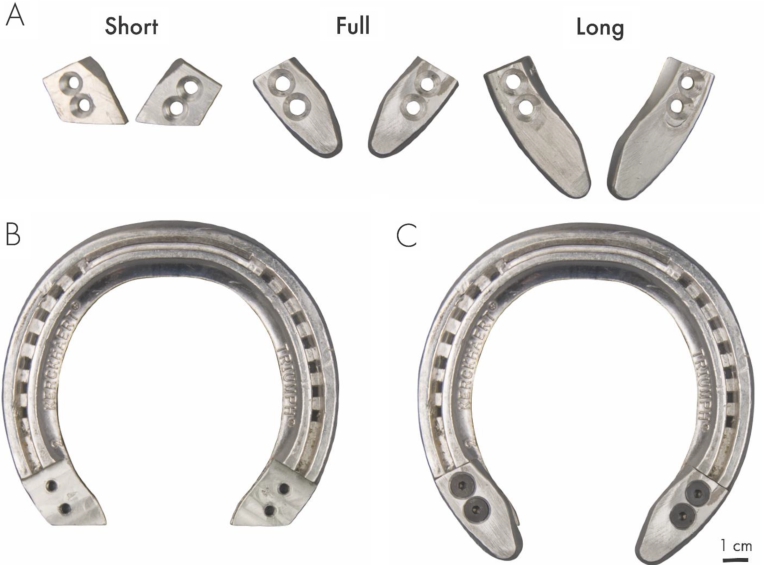
A horseshoe was customised to incorporate the short, full, and long horseshoe lengths (Figure 1A-C). These lengths were defined as the palmar extent of the horseshoe branch aligned with:
- â the widest part of the frog at the heels (Full);
- â 9% shorter than full shoe length, dorsal to the widest part of the frog (Short);
- â 9% longer than full shoe length, palmar to the widest part of the frog (Long).

Figure 1. (A) Shoe branch lengths. (B) Modified shoe without the branch lengths attached showing predrilled holes to screw the branch lengths to. (C) Modified shoe with full pieces that have been screwed into place to show how the shoe was set up with the heel pieces attached.
Citation: Comparative Exercise Physiology 19, 5 (2023) ; 10.1163/17552559-20220071
Limb specimens
Nine unilateral forelimbs from nine riding type horses (median age 16 years, range 6-25 years; 4 females, 4 geldings, 1 unknown; 3 Quarter Horse, 3 Thoroughbred, 1 Appaloosa, 1 Paint, 1 unknown) that were euthanised for reasons unrelated to lameness were removed by transection at the level of the middle of the radius to maintain the passive components of the fetlock stay apparatus. (Kingsbury et al., 1978; Singer et al., 2015) Criteria for limb selection included absence of evidence of orthopaedic disease in either front limb and absence of hoof capsule abnormalities. Limbs were wrapped in water-tight bags and stored frozen at â20 °C to maintain limb and hoof hydration until limbs were thawed to room temperature prior to instrumentation and testing.
Limb preparation
Hooves were trimmed and shoes were applied by a single Certified Journeyman farrier to ensure uniformity in hoof balance. To simulate the hoofâs interaction with substrate, a silicone-based hoof packing material (Sil-Pak, Vettec Hoof Care, Pomona, CA, USA; Stiffness: 2e6 N/m) was applied on the solar surface of the hoof to the level of the weight bearing surface of the hoof wall (NS condition) or shoe. Four nails secured the shoe to the hoof, 2 nails in the toe and 1 nail in each quarter through the manufacturerâs (Aluminum Triumph, The Royal Kerckhaert Horseshoe Company, Rapenburg, the Netherlands) pre-drilled nail holes. To avoid disruption of the hoof wall instrumentation, nails were not clenched to allow for application and removal of the horseshoe after testing the initial NS condition and before the final NS condition after the horseshoe treatments had been completed.
Horseshoe design
Each limb was fitted with a full branch length shoe as defined above. A step was machine cut in the shoe to allow for the different length branches to be rigidly interlocked on the shoe with two screws placed through predrilled and tapped holes in the shoe. Different length branches were interchanged during testing while the shoe template remained affixed to the hoof (Figure 1A-C).
Limb instrumentation
The hoof was instrumented with strain gauges and kinematic markers. Local deformation was measured using rosette strain gauges (VPG, Malvern, PA, USA) rectangular, 45° three-element) attached at six locations: three proximal to distal locations on the quarter (widest part of the hoof) and heel (halfway between the quarter and palmar aspect of the heel) of the lateral hoof wall. Gauges were placed at approximately 25, 50 and 75% of the total distance from the hairline (coronet band) to the hoof solar margin. Proximal to distal locations were proximal heel (PH), middle heel (MH), distal heel (DH), proximal quarter (PQ), middle quarter (MQ) and distal quarter (DQ) (Figure 2A). The middle arm of the rosette gauges was oriented parallel to the longitudinal axis of the horn tubules (Figure 2A). The 90-270° axis of the gauge coordinate system was aligned with the tubules. Strain gauges were attached to a signal conditioning system (SCXI-1520, PCI-MIO-16E4 Labview 7.1, National Instruments Corporation, Austin, TX, USA).
Schematic diagrams for video marker and strain gauge locations. (A) Strain gauge (gauge icon) and kinematic marker (open circles) locations on the lateral aspect of the right forelimb hoof. The coordinate system for strain directions is aligned relative to horn tubules. (B) Kinematic hoof markers were placed at 33 and 66% from the hairline to the solar surface of the hoof for both heel and quarter locations. Regional hoof deformation measurements used chords defined between kinematic hoof markers. (C) Palmar view of the limb in the mechanical testing system instrumented with kinematic bone and hoof markers used for measuring fetlock angle and proximal and distal quarter and heel expansions. PH, MH, and DH refer to proximal, middle, and distal heel locations, respectively. PQ, MQ, and DQ refer to proximal, middle, and distal quarter locations, respectively.
Citation: Comparative Exercise Physiology 19, 5 (2023) ; 10.1163/17552559-20220071
Hoof wall deformation was measured with kinematic markers applied to the medial and lateral aspects of the hoof quarter and heel, placed at 33% and 66% of the distance from the coronet band to the hoof solar margin (Figure 2B). Kinematic markers were spherical balls (3/8â diameter polytetrafluoroethylene balls, McMaster-Carr, Atlanta, GA, USA) covered by silver reflective tape (Scotchlite 3M, St. Paul, MN, USA) attached to 4.8 mm diameter Steinmann pins. Pins were placed approximately 5 mm into the hoof wall avoiding damage to underlying soft tissue structures.
To measure fetlock extension, pins were inserted perpendicular to the long axis of the limb in a lateromedial direction in predrilled 4 mm diameter holes in the proximal and distal quarter of the length of the bone aspects of MC3 and P1 (Figure 2C). Dorso-palmar radiographs (Digital processor: Mark III, Sound-Eklin DR, Carlsbad, CA, USA, Generator: HF100/30+, MinXray, Inc., Northbrook, IL, USA) were taken (58 kVp, 10.2 mAs, 1 meter film-focal distance) of the instrumented limb to allow conversion of kinematic marker positions to MC3 and P1 reference frames to be used later for angle measurements within the Motus software (Motus 9.0, VICON, Centennial, CO, USA).
Mechanical testing
Mechanical loading was performed in a servohydraulic material testing system (Model 662.10A-08, MTS Systems Corporation, Eden Prairie, MN, USA, Model 809; MTS Systems Corp., Minneapolis, MN, USA) equipped with an axial-torsional load transducer (axial load range 220 kN, resolution 22 N, torsional range 2.5 kNm resolution 0.25 Nm) and software (Multipurpose Testware, TestStarII, MTS Systems Corporation). The proximal end of the radius was potted in an aluminium cylinder with polymethylmethacrylate (PMMA, Co Tray Plastics, GC America Inc, Alsip, IL, USA) and secured to the mechanical testing system. The foot was secured to the system actuator via a translation table on linear bearings to allow the hoof to translate dorsally during limb loading which maintained the radius and metacarpal bones parallel to the axis of loading. Initial positioning was standardised by obtaining a physiologic (mean 210°) palmar fetlock angle at a load of â¼700 N. The limb was preconditioned by 200 sinusoidal cycles (700-1,800 N) at 0.25 Hz under axial displacement control with rotation displacement fixed. Subsequently, the limb was similarly loaded (700-6,700 N) for three sinusoidal cycles at 0.25 Hz (â¼3,350 N/s) to capture peak vertical forces for stance (1,800 N), walk (3,600 N), trot (4,600 N) (Kingsbury et al., 1978) and canter (6,700 N) while axial load and displacement data were captured at 60 Hz. Strain data were collected at 20 Hz to allow for multiplexing of the 24 channels. Video images (Photron Fastcam PCI cameras and Fastcam Viewer Photron, San Diego, CA, USA) were acquired at 60 Hz from palmaromedial and palmarolateral positions (S-PRI, AOS Techologies, AG, Dättwil, Switzerland) to capture the positions of kinematic markers in a calibrated 3D field. One limb did not achieve 6,700 N during testing, so 6,200 N was selected as the maximum load for data analysis to compare data at loads that were achievable for all specimens.
Data reduction
Strain measurements from the rosette gauges were reduced to principal strain magnitudes and directions with a custom program in Matlab (R2010a, The Mathworks, Natick, MA, USA) based on equations by Schajer (1990). Orientation of the left limb principal strains were mirrored so that the direction of gauges relative to anatomy matched the right limbs.
Fetlock angle, hoof wall deformations and heel expansion were determined from the positions of kinematic markers using kinematic analysis software (Motus 9.0, VICON). Fetlock extension was measured as the palmar angle between MC3 and P1. Lateral wall deformations were determined for six chord lengths between kinematic markers on the lateral side of the hoof (Figure 2B). These chord distances are defined as: Proximal (Pr), Distal (Di), Dorsal (Do), Palmar (Pa), Proximo-dorsaldistopalmar (PrDo-DiPa) and Proximopalmar-distodorsal (PrPa-DiDo). Quarter and heel expansions were calculated by the change in chord distances between medial and lateral markers for proximal and distal quarter and heel segments (Figure 2C).
All kinematic measurements were calculated as the difference from stance load (1,800 N) to 3,600 N, 4,600 N and 6,200 N loads for the respective variables. Load and kinematic data were down sampled to 20 Hz to match the strain data recording rate. To ensure that data at specific loads were compared, a forecast technique (MS Excel, Microsoft, Redmond, WA, USA) interpolated strain and kinematic data for the desired loads. Linear and angular resolutions of kinematic markers were 0.01 mm and 0.11°.
Statistical analysis
An analysis of variance (Proc Mixed, SAS Institute, Cary, NC, USA) that accounted for repeated measures was used to assess the effects of being barefoot (NS) and shod with shoes of different length (short, full, long) on hoof wall strains, deformations, heel expansion and fetlock angle using a commercial statistical program (SAS). Differences likely related to stress relaxation of tissues with repeated loading, were found for most outcome measurements between initial and final NS treatments, so linear adjustments (Excel) were made to normalise all data for the difference between initial and final no shoe values among the sequential order of treatments. Treatment, load, order, and the treatment à load interaction were treated as fixed effects. Horse was treated as a random effect. The treatment à load interaction was not statistically significant (
The residuals from the analysis of variance (ANOVA) were examined for normality using a Shapiro-Wilks test (Proc Univariate, SAS). The residuals were considered normally distributed if
3 Results
Hoof strains
Principal tensile strains for PH and MH segments decreased while MQ strains increased as loads increased. Principal compressive strains and shear strains increased for all gauges as load increased (Table 1).
Least squared means and standard error for principal tensile, compressive and shear strain magnitudes by load when averaged over all treatments1,2
Citation: Comparative Exercise Physiology 19, 5 (2023) ; 10.1163/17552559-20220071
Principal tensile strains showed no differences among treatments (Table 2, Figure 3A). Principal compressive strains were larger than principal tensile strains in magnitude. Principal compressive strains decreased from the unshod condition with the application of a long shoe, except in the MQ location. Shear strains had the largest increases with application of a shoe in the DQ location and increasing shoe length in the MH and DH locations (Figure 3B). Shear strains did not vary among treatments in the PH and PQ locations (Table 2, Figure 3C).
Least squared means and standard error for principal tensile, compressive and shear strain magnitudes by treatment when averaged over all loads1,2
Citation: Comparative Exercise Physiology 19, 5 (2023) ; 10.1163/17552559-20220071
Strain direction
Principal directions for tensile strains rotated in a clockwise manner in relation to the orientation of the tubules on the lateral wall of the right hoof with increasing load for PH, DH and PQ gauges by 15, 34, and 3°, respectively (Table 3, Figure 2A). For principal compressive and shear strain directions the PH, MH, and PQ rotated clockwise with increasing load by 5, 12, and 3°, respectively, while the DH and DQ rotated counterclockwise by 6 and 14°, respectively (Figure 3B,C).
Principal tensile strains directions rotated counterclockwise by 9° for the MQ gauge with increasing branch length (Table 4, Figure 2A). Principal compressive and shear strain directions rotated in a counterclockwise manner by 9° and 11°, respectively, for the MQ and DQ locations.
The magnitudes and directions of tensile (A), compressive (B) and maximum shear (C) principal strains are illustrated for each location and treatment on the lateral wall of a right hoof. PHÂ = proximal heel, MHÂ = middle heel, DHÂ = distal heel, PQÂ = proximal quarter, MQÂ = middle quarter, DQÂ = distal quarter. Scales are not the same for each panel to allow for visualization of smaller microstrain magnitudes.
Citation: Comparative Exercise Physiology 19, 5 (2023) ; 10.1163/17552559-20220071
Principal strain directions least squared means and standard errors for compressive, tensile and shear principal strains by load when averaged over all treatments1,2
Citation: Comparative Exercise Physiology 19, 5 (2023) ; 10.1163/17552559-20220071
Principal strain directions least squared means and standard errors for principal compressive, tensile and shear strains by treatment when averaged over all loads1,2
Citation: Comparative Exercise Physiology 19, 5 (2023) ; 10.1163/17552559-20220071
Fetlock extension
Fetlock extension increased as load increased (Table 5). Fetlock extension increased with increasing shoe length (
Fetlock angle, hoof expansion, and wall deformation variables (least square means and standard error) by load when averaged over all treatments1,2
Citation: Comparative Exercise Physiology 19, 5 (2023) ; 10.1163/17552559-20220071
Fetlock angle, hoof expansion and wall deformation variables (least square means and standard errors) by treatment when averaged over all loads1,2
Citation: Comparative Exercise Physiology 19, 5 (2023) ; 10.1163/17552559-20220071
Hoof expansion
Hoof expansion increased with increasing load for proximal and distal heel and quarter variables (Table 5). Hoof expansion differences were not detected among shoe lengths (Table 6).
Hoof deformation
Distance between lateral hoof wall segments decreased with increasing loads for Pr, Di, PrDo-DiPa, and PrPa-DiDo variables; and increased with increasing loads for Pa and Do variables (Table 5). Hoof deformations were not detected among any of the shoe length treatments (Table 6).
4 Discussion
The hoof behaved in a manner expected under increasing loading conditions. Fetlock extension increased with increasing in vitro limb load simulating the middle of stance, consistent with in vivo loading in live horses (Harrison et al., 2010). We found that principle compressive hoof wall strains were larger than principal tensile strains during loading, consistent with Thomasonâs strain experiments in the hoof wall when measured on the lateral aspect of the hoof (Thomason et al., 1992). Observed heel and quarter expansion with increased loading is consistent with the findings of Douglas et al. (1996). We found that expansion of the heels was greater than expansion of the quarters. Three factors could have contributed to this finding. Greater expansion at the heels results from the normal hoof anatomy in which the hoof wall is thickest dorsally, gradually thinning and becoming more flexible toward the palmar aspect of the hoof (Bowker, 2003; Lancaster et al., 2013). The hoof capsule is an open truncated cone with the opening at the heels, allowing the heels to move further than the quarters. In addition, the quarter region of the hoof wall could be constrained by horseshoe nails extending to the widest region of the hoof while the heels would be unconstrained. Increased loading caused the lateral aspect of the hoof wall to lengthen in the proximodistal direction and shorten in the dorsopalmar direction. Thomason et al. (1992) modelled hoof behaviour in vivo using rosette strain gauges placed at the toe, medial and lateral quarters and showed that the palmar part of the hoof not only flexes outward at the distal margin of the hoof, but that some concavity occurs along the hoof wall as the distal portion slides outward. In the current study shortening in the dorsopalmar direction could be due to concavity of the lateral wall during hoof loading.
Increasing shoe length resulted in a small increase in fetlock extension (â¤1°). Our hypothesis was that a shorter shoe branch would increase fetlock extension with less support at the heels. Our findings of the longer branch increasing fetlock extension were unexpected but potentially explained from work by Hüppler et al. (2016). Adding hoof heel support with a longer horseshoe branch hoof tends to decrease downward heel rotation in soft surfaces (Hüppler et al., 2016). Although the experimental set-up did not allow alteration of the hoof rotation in a substrate, the lengthening of the horseshoe branch appears to have altered the degree of fetlock extension, potentially altering the biomechanics of the hoof. The longer branch of the shoe was prevented from rotating within a softer surface in the current study as an unyielding surface was used for testing. It is possible that constraint of distal interphalangeal (coffin) joint rotation could account for the slight increase in fetlock extension because fetlock and coffin joint motions are both affected by tension in the deep digital flexor tendon.
Horseshoe length had no statistically significant effect on hoof wall deformations. Horseshoes are fixed to the hoof with nails at locations dorsal to the widest part of the hoof, thus dorsal to all measurement sites in this study. Consequently, changing the length of the shoe palmar to all attachment sites may not affect lateral movement of the heel and quarter portions of the hoof. It is possible that since all shoes were attached in the same manner with toe and quarter nails, there were no changes in how the hoof was allowed to move, indicating that the amount of hoof wall coverage is less important than how the hoof is constrained by nail placement. Nail placement has been found to alter hoof mechanics in that placing nails closer to the heel of the hoof restricts expansion of the heels (Dahl et al., 2023).
Horseshoe length affected the magnitude of principal compressive and shear strains, with no significant alteration in principal tensile strains. Principal compressive strains decreased from NS to a long shoe. This could be due to a more even distribution of forces applied to the hoof wall with increasing ground coverage that occurs with the longer branch length. A more even distribution of pressure between the shoe and hoof around the weight bearing surface of the hoof was found when coverage at the heels was increased with bar shoes (Hüppler et al., 2016). Perhaps the same effect is occurring with the longer branches as with the bar shoe. However, principal shear strains increased in the middle and distal heel locations with increasing horseshoe length.
Changes in principal shear and compressive strain directions with increasing shoe length were observed in the middle and distal quarter regions as well as principal tensile strain direction for the middle quarter. It is possible that the quarters are restricted by horseshoe nails placed in the widest part of the hoof, affecting the change of principal compression and shear strain directions. As movement of quarter of the hoof may have been restricted by the nail, the middle and distal quarter locations closest to the nail region would be constrained as the hoof was loaded and forces distributed more toward the heels as the branches lengthened. Additionally, as load increased, the distal quarter rotated counterclockwise while the rest of the gauges rotated in a clockwise manner which could potentially be explained by proximity to the most palmar nail placed within the quarter of the hoof.
Limitations of the study included the in vitro and short-term nature of the mechanical tests. While findings observed with increasing horseshoe length were relatively small, the effect of the changes in the live horse over months could be larger and result in a clinically relevant change in hoof conformation. While passive structures of the fetlock stay apparatus were maintained in the cadaveric limb preparation, potential muscle contributions to loading of distal tendons were not incorporated in the study. However, passive structures are the largest contribution to maintaining fetlock support during locomotion (Wilson et al., 2001). Motion of hoof wall kinematic markers out of the plane of mediolateral heel and quarter expansion and rotation of pins for lateral wall markers were unaccounted for in the study. Consequently, changes in chord distances could have incorporated linear and rotational transformations. Ultimately increased confidence in our findings would have to be obtained by 3-dimensional measurement systems. Hoof rotation that might occur in natural surfaces was prevented by rigid attachment to a flat metal plate. Repeating the study on more natural surfaces would be important to consider.
In conclusion, under the conditions studied, there is no evidence that horseshoe length affects hoof wall expansion and deformation; however, principal compressive and shear strain magnitude and direction altered with shoe branch length. Shorter shoe lengths did not increase fetlock extension, however, longer branch lengths did increase fetlock extension. Increasing shoe branch length decreases compression along the lateral hoof wall which could indicate a more even distribution of forces along the hoof wall with increased support at the heels. However, the shortening of shoe branch length did not influence hoof mechanics on a flat rigid surface as expected and does not appear to contribute to the development of underrun heels over time. Further studies should be done to see whether the effects of surface type, exercise, and trimming and shoeing frequency with varying shoe lengths change overall hoof conformation over time.
Corresponding author; e-mail:Â vedahl@ucdavis.edu
Acknowledgements
Supported in part by the Center for Equine Health with funds provided by the State of California satellite wagering fund and contributions by private donors as well as funds provided by the Veterinary Orthopedic Society. The authors thank Matthew Seitzler, Shane Westman, Chrisoula Skouritakis, and Dr. Andrew Blandino for technical and statistical assistance.
Authorsâ contribution
All authors contributed equally to the writing of this paper. All authors read and approved the final manuscript.
Conflict of interest
The authors have declared no conflict of interest.
References
Anderson, T.M., McIlwraith, C.W. and Douay, P., 2004. The role of conformation in musculoskeletal problems in the racing Thoroughbred. Equine Veterinary Journal 36: 571-575. https://doi.org/10.2746/0425164044864462
Balch, O.K., Helman, R.G. and Collier, M.A., 2001. Underrun heels and toe-grab length as possible risk factors for catastrophic musculskeletal injuries in Oklahoma racehorses. American Association of Equine Practioners Proceedings 47: 334-338.
Bellenzani, M., Greve, J.M.D. and Pereira, C.A.M., 2007. In vitro assessment of the equine hoof wall strains in flat weight bearing and after heel elevation. Journal of Equine Veterinary Science 27: 475-480. https://doi.org/10.1016/j.jevs.2007.10.003
Bowker, R.M., 2003. The growth and adaptive capabilities of the hoof wall and sole: functional changes in response to stress. In: AAEP, New Orleans, LA. American Association of Equine Practioners, pp. 146-168.
Brunsting, J., Dumoulin, M., Oosterlinck, M., Haspeslagh, M., Lefere, L. and Pille, F., 2019. Can the hoof be shod without limiting the heel movement? A comparative study between barefoot, shoeing with conventional shoes and a split-toe shoe. Veterinary Journal 246: 7-11. https://doi.org/10.1016/j.tvjl.2019.01.012
Butler, D., 1995. The principles of horseshoeing II. Butler Publishing, La Porte, CO, USA, pp. 19-28, 183-195.
Dahl, V.E., Singer, E.R., Garcia, T.C., Hawkins, D.A. and Stover, S.M., 2023. Hoof expansion, deformation, and surface strains vary with horseshoe nail positions. Animals 13: 1872. https://doi.org/10.3390/ani13111872
Douglas, J.E., Mittal, C., Thomason, J.J. and Jofriet, J.C., 1996. The modulus of elasticity of equine hoof wall: implications for the mechanical function of the hoof. Journal of Experimental Biology 199: 1829-1836. https://doi.org/10.1242/jeb.199.8.1829
Eliashar, E., 2012. The biomechanics of the equine foot as it pertains to farriery. Veterinary Clinics of North American Equine Practitioners 28: 283-291. https://doi.org/10.1016/j.cveq.2012.06.001
Floyd, A.M. and Mansmann, R.A., 2007. Equine podiatry. Saunders Elsevier, Philadelphia, PA, USA, pp. 379-392.
Harrison, S.M., Whitton, R.C., Kawcak, C.E., Stover, S.M. and Pandy, M.G., 2010. Relationship between muscle forces, joint loading and utilization of elastic strain energy in equine locomotion. Journal of Experimental Biology 213: 3998-4009. https://doi.org/10.1242/jeb.044545
Hüppler, M., Hafner, F., Geiger, S., Mader, D. and Hager, J., 2016. Modifying the surface of horseshoes: effects of eggbar, heartbar, open toe, and wide toe shoes on the phalangeal alignment, pressure distribution, and the footing pattern. Journal of Equine Veterinary Science 37: 86-97. https://doi.org/10.1016/j.jevs.2015.12.009
Kane, A.J., Stover, S.M., Gardner, I.A., Bock, K.B., Case, J.T., Johnson, B.J., Anderson, M.L., Barr, B.C., Daft, B.M., Kinde, H., Larochelle, D., Moore, J., Mysore, J., Stoltz, J., Woods, L., Read, D.H. and Ardans, A.A., 1998. Hoof size, shape, and balance as possible risk factors for catastrophic musculoskeletal injury of Thoroughbred racehorses. American Journal of Veterinary Research 59: 1545-1552.
Kingsbury, H.B., Quddus, M.A., Rooney, J.R. and Geary, J.E., 1978. A laboratory system for production of flexion rates and forces in the forelimb of the horse. American Journal of Veterinary Research 39: 365-369.
Lancaster, L.S., Bowker, R.M. and Mauer, W.A., 2013. Equine hoof wall tubule density and morphology. Journal of Veterinary Medical Science 75: 773-778. https://doi.org/10.1292/jvms.12-0399
Mansmann, R.A., James, S., Blikslager, A.T. and vom Orde, K., 2010. Long toes in the hind feet and pain in the gluteal region: an observational study of 77 horses. Journal of Equine Veterinary Science 30: 720-726. https://doi.org/10.1016/j.jevs.2010.11.007
Moyer, W., 1980. Corrective shoeing. Veterinary Clinics of North American Large Animal Practioners 2: 3-24.
Schajer, G.S., 1990. An alternate computational approach to strain-gage rosette data reduction. Experimental Techniques 14: 54-57. https://doi.org/10.1111/j.1747-1567.1990.tb01083.x
Singer, E., Garcia, T. and Stover, S., 2015. Hoof position during limb loading affects dorsoproximal bone strains on the equine proximal phalanx. Journal of Biomechanics 48: 1930-1936. https://doi.org/10.1016/j.jbiomech.2015.04.014
Thomason, J.J., Biewener, A.A. and Bertram, J.E.A., 1992. Surface strain on the equine hoof wall in-vivo implications for the material design and functional morphology of the wall. Journal of Experimental Biology 166: 145-168. https://doi.org/10.1242/jeb.166.1.145
Wilson, A.M., McGuigan, M.P., Su, A. and Van den Bogert, A.J., 2001. Horses damp the spring in their step. Nature 414: 895-899. https://doi.org/10.1038/414895a
Yoshihara, E., Takahashi, T., Otsuka, N., Isayama, T., Tomiyama, T., Hiraga, A. and Wada, S., 2010. Heel movement in horses: comparison between glued and nailed horse shoes at different speeds. Equine Veterinary Journal 42 Suppl. 38: 431-435. https://doi.org/10.1111/j.2042-3306.2010.00243.x